General information
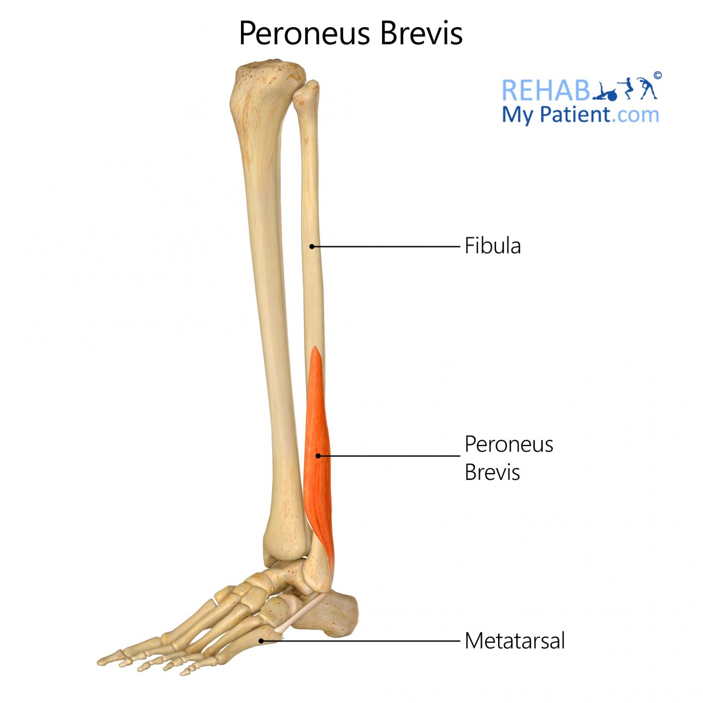
Peroneus brevis is the smaller of two lateral muscles of the leg that assists in plantar flexing and everting the foot and providing lateral stability to the ankle.
Literal meaning
Short muscle of the brooch, or fibula.
Interesting information
Peroneus brevis is attached part way down the fibula running down the outside of the leg and connecting to the bone that lies in front of the fifth metatarsal. Injury often occurs with activities that cause repetitive use of the ankle. Peroneus brevis pain can also be caused by twisting the ankle, crossing legs when seated, and high heels. Treatments for this area includes resting the foot, additional arch support when wearing shoes, wearing a brace to support the area, as well as NSAIDs, such as ibuprofen, to reduce the pain and inflammation.
Origin
Distal 2/3 of the lateral shaft of the fibula.
Insertion
Tuberosity on lateral side of proximal end of 5th metatarsal.
Function
Plantar flexes and everts foot at ankle.
Gives lateral stability to the ankle.
Nerve supply
Superficial fibular (peroneal) nerve (L4, L5, S1).
Blood supply
Muscular branches of the peroneal arteries.

Relevant research
Clinicians investigated the effects of a six-week, multi-station exercise program, integrated into study participants’ normal training programs, on chronic ankle instability. Significant improvements were seen at the conclusion of the study, with subjects reporting reduced frequency of ankle sprains and a better feeling of ankle stability. Multi-station exercises included: exercise mats, Posturomed, ankle disk, Pedalo, exercise band, Air Squab, wooden inversion-eversion boards, mini-trampoline, aerobic step, uneven walkway, Haromed, and Biodex Balance System.
Eils, E., & Rosenbaum, D. I. E. T. E. R. (2001). A multi-station proprioceptive exercise program in patients with ankle instability. Medicine and science in sports and exercise, 33(12), 1991-1998.
Soft tissue and bone variants and abnormalities of the ankle are often detected through the use of magnetic resonance (MR) imaging. A common disease of the ankle associated with pain and instability is a low-lying peroneus brevis muscle. This can predispose one to lateral ankle disease. Radiologists should have knowledge of the MR imaging appearances in order to make precise diagnosis of disorders of the peroneal tendons.
Wang, X. T., Rosenberg, Z. S., Mechlin, M. B., & Schweitzer, M. E. (2005). Normal Variants and Diseases of the Peroneal Tendons and Superior Peroneal Retinaculum: MR Imaging Features1. Radiographics, 25(3), 587-602.
Peroneus brevis exercises
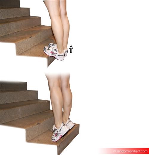
Standing calf raises

Stand on a step with the heels hanging over the edge. Hold on to a wall or handrail for support. Stand tall and pull abdominals in. Bend the knees slightly so the calf muscles aren't fully extended, and then raise heels high. Hold the position for a moment, then lower heels below the edge of the step until a stretch in the calf muscles is felt. Repeat as many at least five times or more, if possible, for three sets. As strength improves, increase the number of repetitions, and also perform the calf raises on one leg at a time.
Balance and reach exercises
Stand next to a chair to provide support, if needed. Stand on one foot and bend the supporting knee slightly. Raise the arch of this foot while keeping the big toe on the floor. Keep foot in this position. With the hand that is farthest away from the chair, reach forward to slightly cross the body, as if to touch the other side of the chair, by bending at the waist. Avoid bending the knee any further while bending. Perform two sets of ten repetitions on each side. To make the exercise more challenging, reach farther forward and down.
Sign Up
Sign up for your free trial now!
Get started with Rehab My Patient today and revolutionize your exercise prescription process for effective rehabilitation.
Start Your 14-Day Free Trial