General information
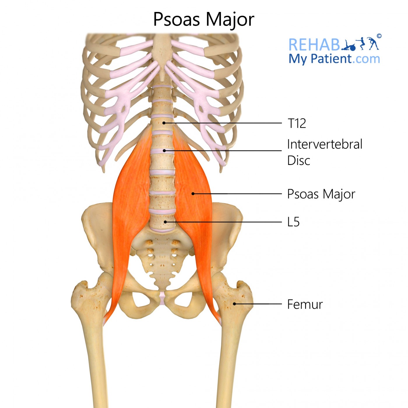
Psoas major is a large muscle located in the posterior abdominal wall that helps move the upper leg and the torso closer together in a flexion movement.
Literal meaning
Greater loin region.
Interesting information
Psoas major is a long, rope-like muscle located on the side of the lumbar region in the vertebral column, running from the spine to the femur. Psoas major forms part of the hip flexors muscle group. It contributes to the flexion and external rotation of the hip joint on the trunk. A common injury is hip flexor strain which occurs due to a sudden contraction or overstretching of the muscle. It often occurs in individuals who participate in sports such as sprinting, football, and rugby. Symptoms include a sudden, sharp burning pain or a tearing and snapping noise during injury. Treatment includes anti-inflammatory medication and painkillers as well as rest, ice, compression, and elevation.
Origin
Transverse processes of lumbar vertebrae; lateral lumbar vertebral bodies.
Insertion
Middle surface of lesser trochanter of femur.
Function
Flexes and laterally rotates thigh at hip.
Flexes vertebral column.
Nerve supply
Anterior primary rami of lumbar plexes L1 – L3.
Blood supply
Lumbar branch of iliolumbar artery.

Relevant research
The purpose of this study was to explore the differences in fibre type composition of the psoas major muscle between its cranial and caudal portion. This study was conducted on 15 males under 35 years old. The types of fibres that compose the psoas major indicate its important dynamic function as the main flexor of this hip joint, its postural function as the stabilizer of the lumbar spine, sacroiliac and hip joints, as well as stabilizing the lumbar lordosis.
Arbanas, J., Starcevic Klasan, G., Nikolic, M., Jerkovic, R., Miljanovic, I., & Malnar, D. (2009). Fibre type composition of the human psoas major muscle with regard to the level of its origin. Journal of anatomy, 215(6), 636-641.
Clinicians studied the evolutionary aspects that may contribute to lower back pain in humans. The most notable feature separating humans from other mammals is the bipedal mode of locomotion. Explanations for lower back pain are hypothesized to be human daily life behaviours, intermuscular coordination, or intramuscular parameters.
Schilling, N., Arnold, D., Wagner, H., & Fischer, M. S. (2005). Evolutionary aspects and muscular properties of the trunk—Implications for human low back pain. Pathophysiology, 12(4), 233-242.
Psoas major exercises

Thomas stretch
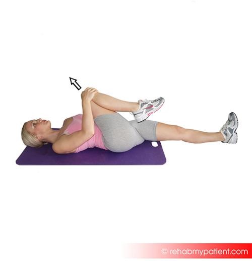
Sit up straight at the end of a padded bench so that the thighs are only halfway on the table. Pull one knee to chest and lean all the way back until lying flat on the bench. The lower back should be flat on the bench. If the back is rounded, or the pelvis is tipped, loosen the hold in order to flatten the back completely. The other leg should hang free off the end of the bench. Hold for 30 to 60 seconds for each side, and complete two to three repetitions.
Kneeling lunge
Kneel on one knee, with the front leg forward at a 90-degree angle. With the hips tucked under, slowly lunge forward, easing into the stretch without straining. Complete 20 reps on each side, holding the lunge for 2 to 3 seconds.
Sign Up
Sign up for your free trial now!
Get started with Rehab My Patient today and revolutionize your exercise prescription process for effective rehabilitation.
Start Your 14-Day Free Trial