General information
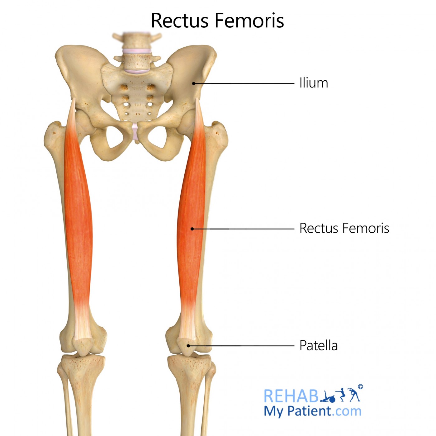
The rectus femoris muscle is one of the four muscles in the quadriceps within the human body. All the components for the quadriceps attach to the patella through the quadriceps tendon. This particular muscle is located along the middle portion of the thigh. Its fusiform shape, superficial fibres arranged in a bipenniform manner and deep fibres running down the deep aponeurosis make this muscle what it is. It serves the function of extending the knee through lifting the lower part of the leg.
Literal meaning
Straight thigh.
Interesting information
The rectus femoris is a quadricep muscle located in the thigh. It crosses along two difference joints: the knee and the hip. Often, it becomes injured as the tendon located at the front of the hip or the muscle within the belly of the thigh. Injuries are referred to as the hip flexor strain. Soccer and football players tend to injure this muscle more often than others. It often involves a forceful movement to cause an injury, like immediately starting to sprint or kicking a ball forcefully, especially when the foot of the athlete hits into another player when trying to kick the ball. Traditional treatments for injuries to this area include rest, icing of the affected area and anti-inflammatory medications to reduce pain and swelling.
Origin
Superior groove in the acetabulum from the anterior surface in the fibrous capsule in the hip joint.
Anterior inferior of the straight tendon.
Insertion
Tibial tuberosity.
Function
Extension of the knee.
Flexion of the hip.
Nerve supply
Femoral nerve L2-L4.
Blood supply
Distal to proximity the muscle is supplied from the deep femoral artery.
Femoral artery.
Descending branch in the lateral circumflex artery.
Genicular anastomoses surrounding the knee.

Relevant research
A 12-week high-intensity exercise program on the composition of fibres and the oxidative capacity found in the rectus femoris muscle from 20 different male rats was determined histochemically. Training exercise program was put into place with a motorized treadmill. The program consisted of four running bouts for two minutes each that alternated with a recovery interval of four minutes. Training helped to increase the relative cross-sectional area within the oxidative fibres and helped to decrease the same parameter within the type IIB fibres that were non-oxidative. Results imply that this type of program is enough to cause changes within the composition of the muscle fibres. It opens the possibility for using this type of exercise to prevent and treat muscle atrophy.
Diaz-Herrera P, García-Castellano JM, Torres A, Morcuende JA, Calbet JA, Sarrat R. Effect of high-intensity running in rectus femoris muscle fiber in rats. J Orthop Res. 2001;19(2):229-232. doi:10.1016/S0736-0266(00)00031-0
Rectus femoris exercises
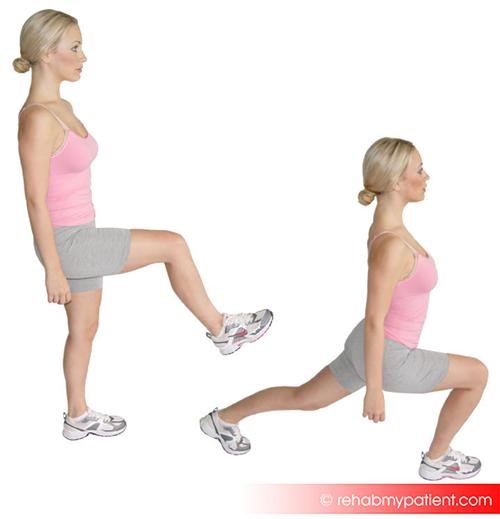
Walking lunges
Begin standing at one end of the room. Step forward with the right foot and bend the legs at the knee to sink to a lunging position. Don’t let the knee extend over the ankle. Push off with the left foot to move forward and meet the other foot. Lead with the left foot and lunge forward. Continue lunging across the room until the other end of the room is reached. Do this for a minimum of twice across the room.

Sign Up
Sign up for your free trial now!
Get started with Rehab My Patient today and revolutionize your exercise prescription process for effective rehabilitation.
Start Your 14-Day Free Trial