General information
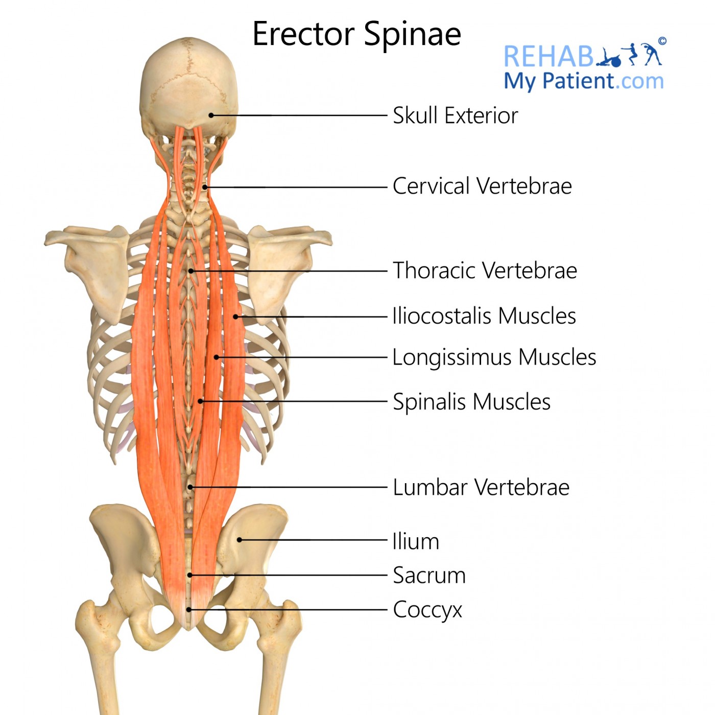
The erector spinae muscles are part of the intrinsic back muscles and form the intermediate layer. The erector spinae are situated superficial to the transversospinales muscles and deep to the serratus posterior superior and inferior muscles. This group consists of three different muscles: the spinalis, longissimus and the iliocostalis. Each of these groups is then further divided and named based on their location along the back.
Literal meaning
Erector of the spine.
Interesting information
The fibres of the erector spinae muscle have a vertical orientation through the cervical, thoracic and lumbar spines and they lie in a groove lateral to the vertebral column. Depending on the region determines what the muscles are covered in, for example the erector spinae in the cervical spine are covered by nuchal ligament, while those in the thoracic and lumbar areas are covered by thoracolumbar fascia.
These muscles are necessary for spinal stability as they assist with posture by stabilising the spine on the pelvis during gait.
Origin
Spinalis capitis: Usually blends with semispinalis capitis
Spinalis cervicis: Spinous process of C7 (sometimes T1 to T2) and ligamentum nuchae
Spinalis thoracis: Spinous process of T11 to L2
Longissimus capitis: C4-T4 transverse process
Longissimus cervicis: T1-T4 transverse process
Longissimus thoracis: Transverse process of lumbar vertebra and blends with iliocostalis in the lumbar region
Iliocostalis cervicis: Angle of ribs 3-6
Iliocostalis thoracis: Angle of lower six ribs
Iliocostalis lumborum: Iliac crest
Insertion
Spinalis capitis: With semispinalis capitis
Spinalis cervicis: Spinous process of C2 and C3-C4
Spinalis thoracis: Spinous process of upper thoracic vertebrae
Longissimus capitis: Posterior edge of the mastoid process
Longissimus cervicis: C2 to C6 transverse process
Longissimus thoracis: Transverse process of all thoracic vertebrae
Iliocostalis cervicis: Transverse process of C4-C6
Iliocostalis thoracis: Angles of upper six ribs and transverse process of C7
Iliocostalis lumborum: L1-L4 lumbar transverse processes, angle of 4-12 ribs and thoracolumbar fascia
Function
Bilateral contraction: Straighten the back, pull the head posteriorly, assist in controlling back flexion.
Unilateral contraction: Side bends/laterally flexes the vertebral column, head rotation.
Nerve supply
Doral rami of spinal nerves.
Blood supply
Branches of the vertebral, deep cervical, occipital, transverse cervical, posterior intercostal, subcostal, lumbar and lateral sacral arteries.
Relevant research
In patients with low back pain there is decreased activity and atrophy of the multifidus muscle which affects spinal stability. The spinal control is then compensated through the increased activity of the erector spinae muscle to stabilize the lumbar spine.
The increased activity of erector spinae increases the compressive load on the vertebral column, which therefore stimulates the nociceptors of the spinal structures continuously which may increase the risk of injury.
Mazis N. Does a history of non-specific low back pain influence electromyographic activity of the erector spinae muscle group during functional movements. J. Nov. Physiother. 2014; 4:226.
Erector spinae exercises
Strengthening:

Roman chair extension
Carefully get onto the Roman chair and start with your head close to the floor and your back in a flexed position. Raise your shoulder up towards the ceiling, extend your back to the horizontal position. Slowly lower yourself again and repeat.
Stretching:

Neck side flexion stretch
Seated, place both hands behind your neck, and gently tilt your neck so that your ear moves closer to your shoulder.
Sign Up
Sign up for your free trial now!
Get started with Rehab My Patient today and revolutionize your exercise prescription process for effective rehabilitation.
Start Your 14-Day Free Trial